Techniques in surgery can be seen as tools to help achieve a specific task. A tennis player’s tools may be seen as his forehand and backhand, his net game and serve, and so on. These tools on their own are not necessarily enough, but rather the right combination of strokes or tools must be achieved in a particular situation.
While a surgeon must master a number of individual techniques for rhinoplasty, he must also have the judgment, skill and natural ability to choose the right techniques for each individual situation.
The different techniques and approaches used in rhinoplasty are detailed below with diagrams and photographs for demonstration purposes.
Taking down the hump:
Cartilage and bone make up the nasal hump. One approach for taking down the hump is described here, but there are several methods that can be used.
To create a guide to visualize the exact amount of hump to be removed, the surgeon may mark the skin of the nasal bridge when the patient is asleep but before local anesthesia is given. Though the amount of reduction is typically determined well before surgery, this provides another helpful guide for the reduction. The surgeon may also have in the operating room the photos of the imaging goal derived from computer imaging to use as another guide.
Dr. Becker’s preferred approach is the en bloc (‘in once piece’) resection of the nasal hump. A scalpel is used for this method to incise the cartilage of the hump in order to create, as shown below, a ‘joint’ at the junction of the cartilage and bone.
As illustrated in the diagrams, the osteotome (bone knife) is situated at the bone-cartilage junction. The osteotome can easily cut through bone because it is extremely sharp. The osteotome advances through the bone in the desired path with a gentle tap-tap technique, then the bone-cartilage hump is removed en bloc, or in one piece.

The bone-cartilage hump that has been removed and the patient’s nose are carefully examined by the surgeon in order to guide the next step which involves additional excision of small amounts of cartilage in order to fine tune the profile and filling or rasping of the bone in order to smooth it. More on rasping is shown below.
Rasping:
A surgical file or rasp is commonly used to reduce or smooth the bony hump. A powered rasp may be used by some surgeons, including Dr. Becker, in order to achieve more precise results. As awareness grows, we hope that more surgeons use the powered rasp. In fact, Dr. Becker has been credited with popularizing the use of powered instrumentation for rasping in rhinoplasty throughout the world.
Osteotomies:
Though not necessary in all rhinoplasties, osteotomies involve ‘cutting’ or ‘breaking’ the nasal bones. Osteotomies are usually required when reducing the nasal hump and are also often used to improve a twisted nose or narrow an overly wide nose.
Though osteotomies may involve several approaches, we have detailed one below:
Medial Osteotomies
Lateral Osteotomies
The bone is cut in the middle with a “back cut” known as a medial osteotomy (diagram on the left). Then, a small bone-knife or osteotome is placed at the edge of the bone as shown. A gentle “tap-tap” technique is used to advance the osteotome along the planned path, outlined in this diagram. Now the bone is cut and may easily be shifted as needed.
For minimally traumatic osteotomies, Dr. Becker has designed small specialty osteotomies which are also available to other surgeons through MicroFrance Corporation, a subsidiary of Medtronic-Xomed. There are many advantages to these “Becker” osteotomies as research has shown that they are less traumatic than the larger, bulkier osteotomies and that less bruising and faster healing times typically result.
Spreader grafts:
Rectangular strips of cartilage known as spreader grafts are placed between the septum and the upper lateral cartilages.
Though fairly uncommon, cases requiring widening of the middle third can benefit from spreader grafts, as shows in this example.


In certain patients, spreader grafts can also provide additional support of the middle third of the nose. An example of that type of patient would be one with short nasal bones, long weak upper lateral cartilages, thin skin and a narrow nose. Another type of patient would be one who requires a large hump reduction and thus needs the additional support of the spreader grafts. An example is shown below:

Suture techniques:
Suture techniques: One reliable yet conservative way to make changes to the nasal tip is through the use of suture techniques. These can be used when refining a bulbous nasal tip or doing a tip elevation (projection and rotation).
Suture techniques are reliable and thus have become increasingly popular. It is believed that these techniques are relatively safe when performed by a competent surgeon because there is no resection of tissue.
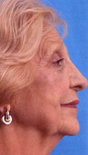
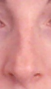
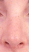
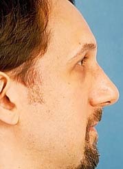
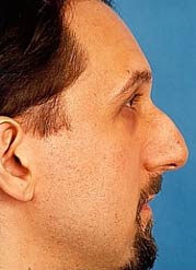
Shown below is a patient who required suture techniques to address a bulbous tip:
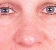
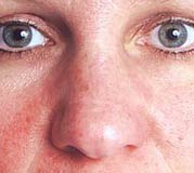

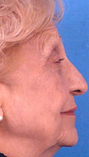
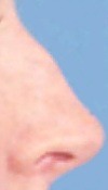
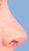
This patient’s nose had a droopy appearance so the tip was lifted (projected and rotated) using suture techniques: