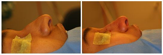
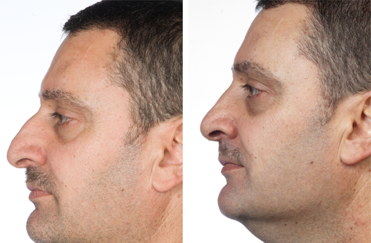
Patient 1
Complaints of a hump, drooping tip and nasal stenosis.
Endonasal approach with septoplasty and hump resection.
Pictures taken in the OR before starting the operation and at the end of the operation to illustrate the tip rotation achiewed from a hump resection.
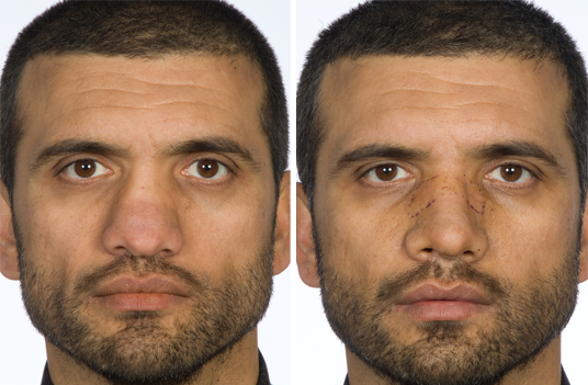
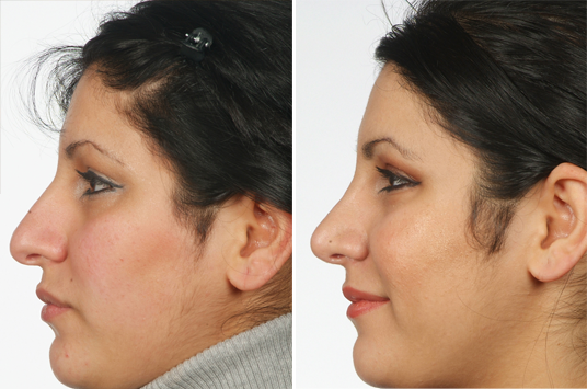
Patient 2
The patient has had a severe nasal trauma. Complaints of broad flat nose with a drooping tip and nasal stenosis. Endonasal approach with septal lenghtening with PDS foil ( see video1 ), cephalic resections ( video 2), osteotomies ( lateral, transversal and paramedian ), separation of upper lateral from septum and dorsal onlay graft.
Pictures: preoperative and one week postoperative.
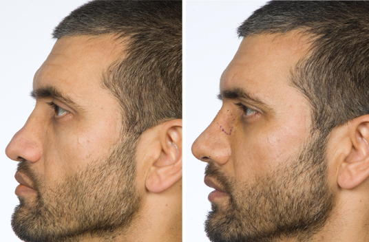
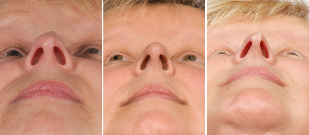
Patient 3
Complaints of a hump, drooping tip, nasal stenosis and too much columella show.
Endonasal approach with septal shortening ( video 3 ), hump resection, cephalic resections ( video 4 ) and resection of returnings of lower margins of upper lateral cartilage ( video 5 ).
Pictures: preoperative and three months postoperative.
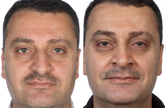
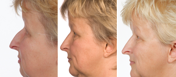
Patient 4
Complaints of a broad and bulbous tip and with hanging infratip columella and hump.
Endonasal approach, hump resection and medial interrupted technique.
Pictures: preoperative and three months postoperative.
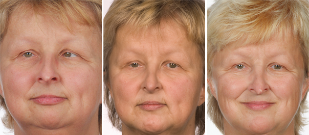
Patient 5
Complaints of a hump and pointed tip with sharp edges.
Endonasal approach, hump resection, delivery with cephalic resections and tip sutures. Still sharp cartilaginous edges.
Revision with medial interrupted technique removes tensions and gives a smooth natural looking tip.
Pictures: preoperative, postoperative after tip sutures and postoperative after medial interrupted technique.
Video 1
Same patient as shown in patient 2.
Cephalic resections performed through transcartilaginous incisions.
( Video 1)
Video 2
Same patient as shown in patient 3.
Video 3
Same patient as shown in patient 2.
Septal lenghtening with PDS foil.
( Video 3)
Video 4
Same patient as shown in patient 3.
Shortening of caudal end of septum, base up triangle.
( Video 4)
Video 5
Medial interrupted strip technique.
( Video 5)
Supplement videos
Video 6
Same patient as shown in patient 3.
Resection of returnings of lower margin of upper lateral cartilage.
( Video 6)
Video 7
Cephalic resection done through a delivery technique.
( Video 7)
The Video-files are DV-PAL anamorphic, interlaced.
Exported from Final Cut Pro.